English
 Français
 Español
 Português
 عربى
عربى 日本語
日本語 한국어
한국어
Suction vs. Nasogastric vs. Feeding Tubes: Use-Case Comparison Guide
Content
Suction tubes, nasogastric (NG) tubes, and feeding tubes are three of the most frequently used enteral devices in clinical settings — yet they are routinely confused, misidentified, or used interchangeably when they should not be. Each device is purpose-built for a distinct clinical role, and selecting the wrong tube can lead to patient discomfort, treatment delays, or serious complications such as aspiration or mucosal damage.
This guide provides a clear, side-by-side comparison of all three tube categories across the dimensions that matter most: clinical use case, tube design, duration of use, material, and procurement considerations. Whether you are a nurse managing bedside care or a hospital procurement manager sourcing disposables at scale, this reference will help you choose the right tube for the right patient at the right time.
What Are Suction, Nasogastric, and Feeding Tubes?
Before comparing use cases, it helps to understand what each device is designed to do at a fundamental level.
Suction catheters are flexible, single- or multi-lumen tubes designed to remove secretions, fluids, or gas from the airway or gastrointestinal tract. In respiratory care, suction catheters are advanced through an endotracheal or tracheostomy tube to clear mucus from the lower airway. In GI care, they are used to decompress the stomach by drawing out accumulated fluids and gas — a function critical after surgery or in cases of bowel obstruction.
Nasogastric tubes are inserted through the nostril, pass through the nasopharynx and esophagus, and terminate in the stomach. The NG tube is a dual-purpose device: it can be used for gastric suctioning (decompression) or for delivering nutrition and medication into the stomach. The tube selected depends entirely on which function is required — suctioning demands a large-bore double-lumen design, while feeding requires a narrow, flexible single-lumen tube.
Feeding tubes encompass a broader category of enteral access devices used when a patient cannot meet nutritional needs orally. Feeding tubes range from short-term nasogastric feeding tubes to surgically or endoscopically placed percutaneous devices such as gastrostomy (PEG) or jejunostomy (PEJ) tubes, which are intended for long-term nutritional support lasting months to years.
Use-Case Comparison: When to Use Which Tube
The table below summarizes the key decision-making criteria when selecting between these three device categories. Clinical context — particularly the intended function and anticipated duration of use — should always drive the choice.
| Criteria | Suction Catheter | Nasogastric (NG) Tube | Feeding Tube (Enteral) |
|---|---|---|---|
| Primary function | Remove secretions or decompress stomach/airway | Gastric decompression or short-term feeding/medication delivery | Nutritional support and medication delivery |
| Insertion route | Nasal/oral (GI) or via endotracheal/tracheostomy tube (airway) | Nasal → esophagus → stomach | Nasal (short-term) or percutaneous via abdominal wall (long-term) |
| Tip location | Stomach (GI suction) or lower airway (respiratory) | Stomach (or post-pyloric for reduced aspiration risk) | Stomach (gastrostomy) or small bowel (jejunostomy/post-pyloric) |
| Duration of use | Intermittent / acute (minutes to hours) | Short-term (typically <4 weeks) | Short-term (NG) to long-term (months–years for PEG/PEJ) |
| Lumen design | Single or double lumen | Double lumen (decompression) or single lumen (feeding) | Single lumen, small bore |
| Bore size | 6–22 Fr (varies by application) | 12–18 Fr (decompression); 8–12 Fr (feeding) | 5–12 Fr (nasal); larger for PEG/PEJ |
| Typical clinical settings | ICU, OR, emergency, post-op recovery | Acute care, emergency, post-op, short-term recovery | Long-term care, home care, neurology, oncology, geriatrics |
| Key indications | Bowel obstruction, ileus, post-anesthesia, airway secretion clearance | Dysphagia, altered consciousness, pre-/post-surgery, GI bleeding evaluation | Neurological disorders, head/neck cancer, malnutrition, chronic dysphagia |
One important clinical consideration is aspiration risk. For patients in whom aspiration is a major concern, post-pyloric placement — advancing the tube tip past the stomach and into the duodenum or jejunum — significantly reduces the likelihood of gastric content entering the lungs. This applies to both NG feeding tubes and dedicated enteral feeding tubes.
Tube Types and Design Differences
Within each category, tube design varies substantially depending on the intended clinical task. Understanding the main models in clinical circulation helps procurement teams order the correct SKU and helps clinicians request the appropriate device.
Salem Sump (double-lumen NG tube): The standard tube for gastric decompression. It features a large lumen for suctioning and a smaller sump lumen that introduces ambient air, preventing the tube from adhering to the gastric mucosa and causing tissue damage during continuous suction. The Salem Sump is typically 14–18 Fr and is connected to a wall suction unit at low intermittent settings (40–80 mmHg).
Levin Tube (single-lumen NG tube): A simpler, smaller-diameter single-lumen tube used for feeding or medication delivery. Without a sump lumen, it is not suitable for continuous gastric decompression, as negative pressure can draw the tube against the stomach wall. It is commonly used for short-term enteral feeding when patient tolerance is a priority.
Dobhoff Tube (weighted feeding tube): A small-bore single-lumen tube with a tungsten weight at the distal tip. The weight is intended to allow peristalsis and gravity to advance the tube past the pylorus into the duodenum, reducing aspiration risk. Dobhoff tubes are preferred when post-pyloric feeding is clinically indicated but endoscopic placement is not immediately available.
PEG and PEJ Tubes: Percutaneous endoscopic gastrostomy and jejunostomy tubes are placed through the abdominal wall under endoscopic or radiologic guidance. They are the preferred choice when nutritional support is expected to exceed four to six weeks. These tubes accommodate larger bore sizes, support more viscous formulas, and substantially improve patient comfort by eliminating the nasal component entirely.
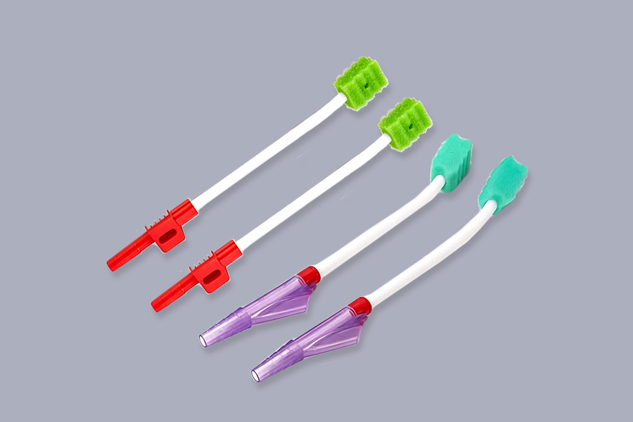
Suction Catheters (airway and GI): Airway suction catheters are thin, flexible, color-coded devices (6–14 Fr for adults) passed through an endotracheal or tracheostomy tube to clear lower airway secretions. GI suction catheters, by contrast, are larger and are used for gastric or intestinal drainage. A related device, the mucus extractor, is specifically designed for upper airway secretion removal in neonates and adult patients unable to clear secretions independently — commonly encountered in ICU and neonatal care units.
Material Considerations: PVC vs. Silicone
The two materials used in the manufacture of these tubes — polyvinyl chloride (PVC) and silicone — carry meaningfully different properties that should factor into procurement and clinical selection decisions.
PVC is the most widely used material for short-term applications. It is cost-effective, less prone to kinking during insertion, and provides sufficient rigidity to ease placement. However, PVC can become stiffer at lower temperatures, and prolonged contact with mucosal tissue increases the risk of irritation or erosion. For tubes expected to remain in place for seven days or fewer, PVC is generally appropriate.
Silicone is the preferred material for longer-duration applications, particularly in pediatric patients or immunocompromised individuals. Medical-grade silicone is highly biocompatible, minimizes allergic reactions, and remains flexible at body temperature without softening excessively. It tolerates high-heat sterilization (autoclaving) without structural degradation, making it more suitable for reprocessable applications. Silicone feeding tubes often incorporate multiple side holes at the distal end to prevent formula clogging and ensure consistent nutrient flow.
Polyurethane represents a third option gaining traction for nasogastric feeding tubes specifically. Polyurethane tubing softens at body temperature while maintaining insertion stiffness at room temperature, striking a balance between ease of placement and long-term patient comfort.
For bulk procurement, the clinical use case should determine the material specification. Acute care wards cycling through tubes frequently may find PVC most economical; long-term care facilities and neonatal units should prioritize silicone to reduce complication rates and tube replacement frequency.
Placement Verification and Key Complications
Regardless of tube type, placement verification is a non-negotiable step. An incorrectly placed tube — particularly one inadvertently advanced into the trachea or lungs — can cause fatal complications including aspiration pneumonia, pneumothorax, or pleural effusion.
Chest or abdominal X-ray remains the gold standard for confirming initial tube placement. It provides definitive visualization of the tube tip location relative to the carina and stomach. For feeding tubes specifically, X-ray confirmation before the first feed is considered mandatory in most clinical guidelines.
Aspirate pH testing offers a practical method for ongoing placement checks in between X-rays. Gastric fluid aspirated through the tube should register a pH of 5.5 or lower, indicating the presence of stomach acid. A higher pH may suggest bronchial or esophageal placement and warrants immediate cessation of any infusion and further verification.
Point-of-care ultrasound (POCUS) is an emerging alternative in ICU settings, offering real-time tube visualization without radiation exposure. Its adoption is growing, particularly in COVID-19 ICU environments where mobility of imaging equipment is limited.
Common complications associated with nasogastric and feeding tube use include mucosal irritation of the nares, sinusitis, epistaxis, and — in serious cases — esophageal or gastric perforation. For suction catheters, over-aggressive suctioning (pressures exceeding 80 mmHg) can cause trauma to the tracheal or gastric mucosa. Prolonged gastric suctioning through an NG tube risks erosion of the gastric lining and electrolyte imbalances from continuous fluid removal.
Choosing the Right Tube for Your Facility
The decision framework is straightforward once the clinical context is clear:
- If the goal is airway or gastric decompression in an acute or surgical setting, a double-lumen NG suction tube (e.g., Salem Sump) or a dedicated airway suction catheter is the correct choice.
- If the goal is short-term enteral feeding or medication administration (under four weeks) for a patient with a functional GI tract, a small-bore single-lumen NG feeding tube — such as a Levin or Dobhoff — is appropriate.
- If the patient requires long-term nutritional support beyond four to six weeks, a percutaneous device (PEG or PEJ) should be considered for improved patient comfort, lower dislodgement risk, and reduced nasal complications.
- If the patient is in the ICU or neonatal unit and requires secretion clearance from the upper airway, a dedicated mucus extractor is the indicated device.
For hospital procurement teams, establishing clear par levels for each tube category — categorized by French size, material, and lumen configuration — reduces bedside decision fatigue and minimizes the risk of substituting one tube type for another under time pressure. Specifying CE- and ISO 13485-certified, single-use, ETO-sterilized products ensures both regulatory compliance and patient safety across all tube categories.